|
III.
Blastomycocis
|
|
Case 23:
Blastomycocis / Myelogenic leukaemia
|
|
|
|
Blastomycocis
|
|
Myelogenic leukaemia
|
|
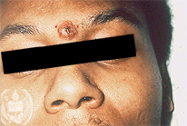
Fig.23-A1
Blastomycosis
A patient from Louisville /Kentucky, USA shows in his face and on his neck several subcutaneous nodules with crusts on the surface. The patient looks healthy. The x-ray examination of his thorax reveals in the upper fields of both lungs numerous infiltrates. After the histological diagnosis a therapy with Ketokonazol was successful.
|
|
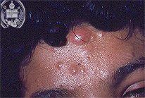
Fig.23-B1
Myelogenic leukaemia
On the forehead of the patient some smaller and one major tumor formation can be seen.
|
|
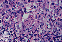
Fig.23-A2
Blastomycosis
The tissue reaction in the HE stain and higher magnification consists in a non-specific granulation tissue with some giant cells. Furthermore, numerous relatively large yeast-like cells are visible laying partly within giant cells and presenting the aspect of double membranes.
|
|
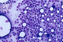
Fig.23-B2
Myelogenic leukaemia
The histology of the bone marrow shows mainly immature white blood cells. Pappenheim stain; 1000 x.
|
|
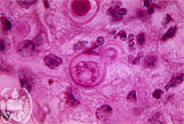
Fig.23-A3
Blastomycosis
With the higher power and in the HE stain large cells of Blastomyces dermatitidis are seen, with solitary budding where the smaller daughter cell is sitting broad on the mother cell. Again the "shrinking aspect" of the membrane can be recognized.
|
|
|
| español | english | deutsch |
|
|