|
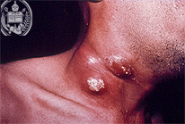
Fig.22-A1
Blastomycosis
Several necrotic skin nodules are present on this patient. They are partly ulcerated and are found on the right side of his neck. The surrounding skin shows a heavy reddening. The causative agent is a dimorphic fungus ocurring in the tissue as a relatively big yeast cell and may reach a diameter up to 23 µ. This deep mycosis occurs by inhalation with primary infection in the lungs as in many other deep mycoses.
|
|
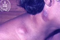
Fig.22-B1
Lymph node tuberculosis
The subcutaneous nodules at the neck of this adolescent patient are swollen lymph nodes. They are not ulcerated. This is a primary form of tuberculosis, found mainly in adolescents, later accompanied by necrosis and formation of fistulae.
|
|
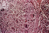
Fig.22-A2
Blastomycosis
The tissue reaction in this mycosis is "mixed", i.e. a non-specific granulation tissue and a granulomatous inflammation with giant cells are found. Frequently also granulomas are observed. In this picture the yeast-like cells in the HE stain and in this magnification are hardly seen.
|
|
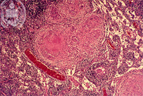
Fig.22-B2
Lymph node tuberculosis
In the lymphatic tissue typical tuberculoses granulomas with central necroses are seen in this HE stain and with low power.
|
|
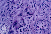
Fig.22-A3
Blastomycosis
With higher magnification and in the HE stain a singular budding is seen in the tissue which represents the multiplication of the fungus. Here mother and daughter cell have the same size. Both fungus cells show the aspect of a double cell membrane, but this is an artifice, because a retraction took place in the preparation of the histological slide. All yeast-like fungus cells have only one membrane.
|
|
|