Begin  The Authors
 Introduction

Tropical Diseases:

I Leishmaniasis
II Paracoccidioidomycosis
III Blastomycocis
IV Chromoblastomycosis
V Lepra
VI Sporotrichosis
VII Histoplasmosis
VIII Rhinosporidiosis
IX Mycetomas
X Rhinoscleroma
XI Chagasdisease
XII Actinomycosis
XIII Mucormycosis
XIV Amoebiasis
XV Protothecosis
XVI Lobomycosis
XVII Phaeohyphomycosis
XVIII Pruritus actinicus
XIX Bite of snake
XX Coccidioidomycosis

 List of cases

 References
 Contact

III. Blastomycocis

Case 22. Blastomycocis / Lymph node tuberculosis

View Thumbnails

Fig. 22-A3

Previus

Fig. 22-A3
Fig. 22-A3: Blastomycosis

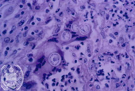
With higher magnification and in the HE stain a singular budding is seen in the tissue which represents the multiplication of the fungus. Here mother and daughter cell have the same size. Both fungus cells show the aspect of a double cell membrane, but this is an artifice, because a retraction took place in the preparation of the histological slide. All yeast-like fungus cells have only one membrane.

Next


| español | english | deutsch |


Facultad de Medicina
Universidad de Los Andes
Merida - Venezuela