|
I.
Leishmaniasis
|
|
Case 9:
Tegumentary Leishmaniasis / Prurigo de Hebra
|
|
|
|
Tegumentary Leishmaniasis
|
|
Prurigo de Hebra
|
|

Fig.9-A1
Tegumentary Leishmaniasis
A farmer boy lived under very poor conditions and developed in the course of several months numerous wart-like skin nodules mostly in the face extending to the neck. The patient was in a very bad shape and had a severe anemia.
|
|

Fig.9-B1
Prurigo de Hebra
A 9-year old boy presented a generalized very itching exanthema of the skin. The disease began 6 months earlier in the face with the appearance of papules which transformed later into purulent pustules extending to the trunk and the extremities.
|
|

Fig.9-A2
Tegumentary Leishmaniasis
In the biopsies of several skin nodules numerous histiocytes with clear cytoplasm were found at low power and in the HE stain. At this magnification intracellular parasites are difficult to recognize. The Grocott stain was negative.
|
|

Fig.9-B2
Prurigo de Hebra
The skin biopsy shows an irregular acanthosis and hyperkeratosis of the epidermis in addition to crusted ulcers. In the deeper layers of the corium extensive inflammatory infiltrates are seen of leuco-and lymphocytes, monocytes and a mayor number of eosinophils. They extend deep to the subcutaneous fat tissue.
|
|
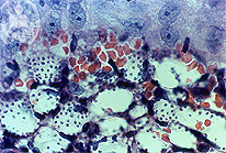
Fig.9-A3
Tegumentary Leishmaniasis
At high power the small granular parasites within the vacuolated histiocytes are good to see. They are leishmanias and are Grocott negative. This form of Leishmaniasis with numerous skin nodules and parasites is a opportunistic infection in of an immunodeficiency syndrome.
|
|
|
| español | english | deutsch |
|
|