|
VIII.
Rinosporidiosis
|
|
Case 53:
Rinosporidiosis / Pyogenic Granuloma
|
|
|
|
Rinosporidiosis
|
|
Pyogenic Granuloma
|
|
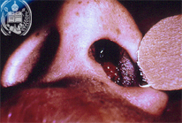
Fig.53-A1
Rhinosporidiosis
A 8-year old boy from the State Barinas / Venezuela shows a small tumor of his left nostril which was noted for the first time several weeks ago. It is painless, but easily bleeding. The tumor has a smooth, dark red surface with little white spots. Clinically, a benign polyp was assumed.
|
|
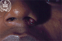
Fig.53-B1
Pyogenic Granuloma
This patient presents in his left nostril a roundish tumor with a diameter of one cm and a smooth surface. This 14 - years old boy developed this nodule 2 weeks ago.
|
|
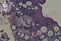
Fig.53-A2
Rhinosporidiosis
In the biopsy the two types of the fungal cells of Rhinosporidium seeberi, the trophic form and several sporangia con be distinguished in the HE stain and at low power. The latter are cystic and contain countless endospores. The fungus cells are easily recognized. A Grocott stain is not necessary.
|
|
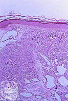
Fig.53-B2
Pyogenic Granuloma
Histologically this tumor showed a flattened epithelium and a non-specific granulation tissue with numerous capillaries and a small number of lymphocytes. Fungi or parasites could not be found, HE stain, higher power.
|
|
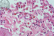
Fig.53-A3
Rhinosporidiosis
After the rupture of the sporangium wall the endospores reach the surface of the mucosa. HE stain; higher power.
|
|
|
|
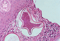
Fig.53-A4
Rhinosporidiosis
After the ejection of the mature endospores a deformation of the sporangia takes place. Later, see 48, foreign body granulomas with giant cells appear. When the fungus cells are intact hardly a tissue reaction is seen. HE stain, higher power.
|
|
|
| español | english | deutsch |
|
|