|
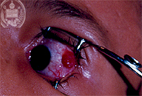
Fig.54-A1
Rhinosporidiosis
A 10-years old girl from the State of Barinas / Venezuela presents in her left eye a roundish, dark red coloured tumor in the conjunctiva, surrounded by hyperaemic vessels. The tumor has a diameter of one cm and a thickness of 2 mm. Clinically, a papilloma of the conjunctiva is assumed. This fungal infection, mostly occurring as a tumor-like lesion which clinically is diagnosed frequently as a polyp or papilloma before the fungal character of the lesion is confirmed histologically. The flatlands of Venezuela (the so called Llanos) with their big rivers are an endemic region for this fungal infection. It occurs almost never in the mountainous zones of the country.
|
|
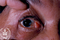
Fig.54-B1
Dermoid
The yellowish -reddish tumor of the conjunctiva is benign. It developed slowly in the course of a long time, but enlarged more especially in the last years.
|
|
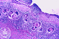
Fig.54-A2
Rhinosporidiosis
Histologically a typical chronic Rhinosporidiosis with numerous giant cells is found wich have phagocytized fungal cells.
|
|
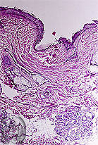
Fig.54-B2
Dermoid
Under the atrophic squamous epithelial cells of the conjunctiva the formation of a tumor is found histologically consisting of connective and fatty tissue with some hair follicles and sebaceous and sweat glands. HE stain; low power.
|