|
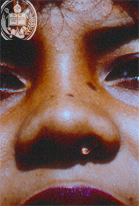
Fig.52-A1
Rhinosporidiosis
In the left nostril of this female patient a small tumor can be seen.
|
|
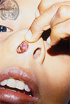
Fig.52-B1
Angiomatous Fibroma
A 9-year old boy presents a painless tumor in his right nostril.
|
|
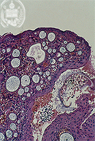
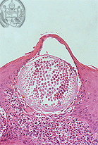
Fig.52-A2
Rhinosporidiosis
With low power and in the HE stain numerous clear trophic cells of R. seeberi showing different sizes are observed. In some of them nuclei are present. Furthermore, a ruptured sporangium with endospores outside of the large fungus cell can be seen.
|
|
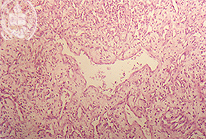
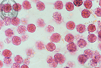
Fig.52-B2
Angiomatous Fibroma
The polipous tumor microscopically consists of connective tissue with abundant cavernous blood vessels.
|