|

Fig.68-A1
Actinic Pruritus
In a 8-year old boy from the flatlands around the Apure river, the so called "Llanos occidentales" developed in the course of several weeks eztensive papulous-vesicular, very itching skin alterations. Especially the forehead, the environs of both eyes, the skin above the cheek bones and the nose were affected. The chin was less affected, but both ears showed vesicular-papillomatous skin lesions. A pharmacodermatitis could be excluded.
|
|
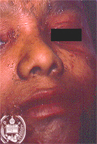
Fig.68-B1
Staphylococcal scalded skin syndrome
Another 8-years old boy felt ill suddenly with high fever, loss of appetite and somnolencia, accompanied by skin lesions, especially in the face. They consisted of superficial blisters whch scaled of easily from the base. They were found mainly on the forehead, the environs of the left eye, the perioral region and the ears.
|
|
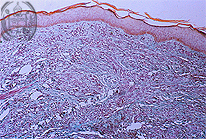
Fig.68-A2
Actinic Pruritus
Histologically there was a diffuse hyperkeratoris of the epidermis. The corium showed an oedema and dense round cell perivascular infiltrates.
|
|
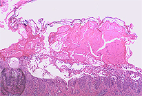
Fig.68-B2
Staphylococcal scalded skin syndrome
Histologically the intraepithelial blister formation containing fibrine and leucocytes could be observed.
|
|

Fig.68-A3
Actinic Pruritus
With high power the dense perivascular infiltrates and the oedema are good to see.
|
|
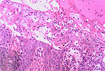
Fig.68-B3
Staphylococcal scalded skin syndrome
With higher power numerous inflammatory elements could be recognized, mostly leucocytes, masses of fibrine and several acantholytic epithelial cells.
|
|

Fig.68-A4
Actinic Pruritus
After the treatment with chloroquine and cortisone the papulous skin lesions transformed into elevated, dark red, and firm plaques which later healed completely.
|
|
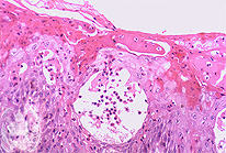
Fig.68-B4
Staphylococcal scalded skin syndrome
Occasionally an intraepithelial blister formation is seen with an exsudate and several acantholytic epithelial cells. The disease is called "Staphylococcal scalded skin syndrome" or "Lyell syndrome".
|