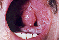
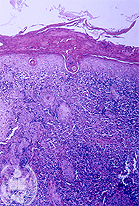
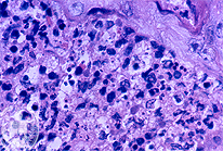
Begin  The Authors
 Introduction

Tropical Diseases:

I Leishmaniasis
II Paracoccidioidomycosis
III Blastomycocis
IV Chromoblastomycosis
V Lepra
VI Sporotrichosis
VII Histoplasmosis
VIII Rhinosporidiosis
IX Mycetomas
X Rhinoscleroma
XI Chagasdisease
XII Actinomycosis
XIII Mucormycosis
XIV Amoebiasis
XV Protothecosis
XVI Lobomycosis
XVII Phaeohyphomycosis
XVIII Pruritus actinicus
XIX Bite of snake
XX Coccidioidomycosis

 List of cases

 References
 Contact

I. Leishmaniasis

Case 11: Mucocutaneous Leishmaniasis / Secondary Syphilis

Previus Case 10

| español | english | deutsch |

Mucocutaneous Leishmaniasis

Secondary Syphilis

Click here to see the picture 11-A1
Fig.11-A1
Mucocutaneous Leishmaniasis

A 27 -years old man from Socopó, a town in the West of the State Barinas comes to an Ear-Nose-Throat specialist presenting nasal ardor and bleeding together with a mucous secretion and hoarseness. The patient had been treated with several antibiotics without any improvement. Looking at the mucosa of the oral cavity a marked diffuse reddening is found, accompanied by several slightly elevated foci involving the palatal velum, the uvula and the soft palate.
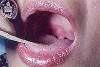
Click here to see the picture 11-B1
Fig.11-B1
Secondary Syphilis

A 23 years -old woman from Barinas showed a painful inflammation of the palate, preferably of the uvula which existed only since a few days. The patient complained of headaches, fever and nausea. In smears taken from the affected mucosa neither fungi nor parasites were found. In the lymph of the mucosal areas and from the lymphnodes moving Trepanoma pallidum organisms could be seen in the darkfield. Furthermore the VDRL test was positive. The patient reported later a small painless ulcer at the introituses vagina 10 weeks ago which had healed spontaneously. Therefore the diagnosis of a secondary syphilitic pharyngitis, so called "plaque muqueuse", could be made.

Click here to see the picture 11-A2
Fig.11-A2
Mucocutaneous Leishmaniasis

In the biopsy a non-specific granulation tissue was found under the epithelium of the oral mucosa with numerous histiocytes and isolated leishmanias. The latter were detected only after a long search. HE stains.

Click here to see the picture 11-A3
Fig.11-A3
Mucocutaneous Leishmaniasis

With the higher magnification the parasites are better to recognize. The absence of leucocytes in the granulation tissue in these cases is typical. The Grocott stain which must be applied in such cases was negative, meaning that no fungal infection existed.


| español | english | deutsch |


Facultad de Medicina
Universidad de Los Andes
Merida - Venezuela