|
IX.
Mycetomas
|
|
Case 57:
Actinomycetoma / Melanoma
|
|
|
|
Actinomycetoma
|
|
Melanoma
|
|
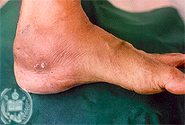
Fig.57-A1
Actinomycetoma
On the plantar surface of the patients foot of this case developed a roundish, poorly limited tumor of a diameter of several cms. Here it may be noted that the three types of mycetomas (eumycetomas, schizomycetomas and actinomycetomas) macroscopically can not be differentiated, because all the three types have similar morphologic characteristics.
|
|

Fig.57-B1
Melanoma
As in 57-A1 a tumor is present on the lateral posterior plantar surface. It is not sharply limited, but looks similar to the actinomycetoma.
|
|

Fig.57-A2
Actinomycetoma
In this case the grains are very irregular and present a pale blue colour in the HE stain. The germs of these grains are nocardiae. In addition, actinomycetes may be the causative agents situated in the grains. As mentioned earlier these germs earlier were classified as fungi, but today as bacteria.
|
|

Fig.57-B2
Melanoma
Histologically an amelanotic melanoma is diagnosed.
|
|

Fig.57-A3
Actinomycetoma
With the Grocott method also these grains stain black. The picture is from the same slide as in the fig. 57-A2 and shows that several grains are present.
|
|
|
|
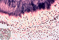
Fig.57-A4
Actinomycetoma
In the periphery of a large grain very small protrusions are visible on the surface representing the filamentous nocardiae.
|
|
|
|
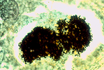
Fig.57-A5
Actinomycetoma
A large grain of the same case is also stained with the Grocott method showing also minimal superficial protrusions of the germs.
|
|
|
| español | english | deutsch |
|
|