|
VII.
Histoplasmosis
|
|
Case 50:
Histoplasmosis / Liposarcoma
|
|
|
|
Histoplasmosis
|
|
Liposarcoma
|
|
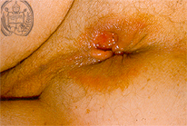
Fig.50-A1
Histoplasmosis
In a female patient hospitalized in Mérida / Venezuela for another disease, accidentally a perianal redness of the skin with small tumor-like nodules was found. The biopsy of the red skin with the small nodules resulted as a typical histoplasmosis. Unfortunately no other clinical dates of this are known. We present this here in order to compare it with another one where a liposarcoma of the right gluteal region was found. At this opportunity we will use the space to present the so-called "morning stars" which occur typically as macroconidias with radial proliferations together with thin hyphae in the culture of the mycelial form of H. caps. var. capsulatum at room temperature. At the contrary, the yeast form of the histoplasmas occurs in tissues of humans and animals at higher temperatures. These two forms are the reason to call this fungus "dimorphic". Furthermore we show two more pictures of the structure of fungi from the other variety of the genus Histoplasma capsulatm, the H.caps. var. duboisii.
|
|
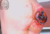
Fig.50-B1
Liposarcoma
This haemorrhagic tumor is localized in the gluteal region of this patient.
|
|
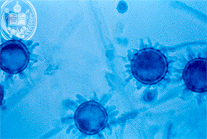
Fig.50-A2
Histoplasmosis
The "morning stars" mentioned are called this way in German, because they are similar to weapons used by the mercenaries in the Middle Ages.
|
|
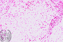
Fig.50-B2
Lipocarcoma
Histologically a liposarcoma could be confirmed.
|
|

Fig.50-A3
Histoplasmosis
A "morning star" by electron microscopy.
|
|
|
|
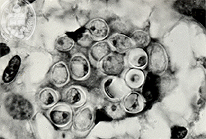
Fig.50-A4
Histoplasmosis (Variedad duboisii)
With the HE stain and high power the large histoplamas are seen, containing nuclei.
|
|
|
|
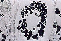
Fig.50-A5
Histoplasmosis (Variedad duboisii)
Numerous large fungus cells of this variety are situated often within giant cells, demonstratd here in the Grocott stain.
|
|
|
| español | english | deutsch |
|
|