|
VII.
Histoplasmosis
|
|
Case 49:
Histoplasmosis / Purulent folliculitis
|
|
|
|
Histoplasmosis
|
|
Purulent folliculitis
|
|
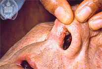
Fig.49-A1
Histoplasmosis
A 64-years old woman from the environs of the town Barinas / Venezuela refers a superficial ulceration of the mucosa of the left nostril existing several months. The patient seems to be healthy. The x-ray picture of the lungs shows a dissemination of small calcified and several bronchopneumonic foci. After the biopsy diagnosis of the nasal mucosa a specific treatment with Amphotericin B was started. About the further evolution of this infection nothing is known.
|
|
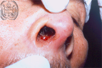
Fig.49-B1
Purulent folliculitis
In the right nostril of another patient developed a lentil-like tumor with a yellowish zone which resembled an infection with fungi. The 62.-year old patient was hospitalized for treatment of a prostatic hypertrophy.
|
|
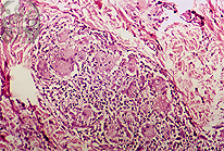
Fig.49-A2
Histoplasmosis
In the biopsy of the nasal mucosa granulomas with giant cells are seen with the HE stain. With this low power fungal elements are hardly observed within the histiocytes.
|
|
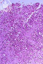
Fig.49-B2
Purulent folliculitis
In addition to a small abscess was found histologically a non specific granulation tissue with histiocytes which had a foamy cytoplasm. Special fungal stains were negative.
|
|
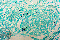
Fig.49-A3
Histoplasmosis
In the granulomas shown in fig. 49-A2, however are found a few small black coloured yeast -like cells, partly situated inside of giant cells are seen with the Grocott stain. Single budding can not be observed with this magnification.
|
|
|
|

Fig.49-A4
Histoplasmosis
In addition to the images of the human we show here a slide from the intestinal mucosa of a bat, which was caught in a cave of the Venezuelan Andes. In this region histoplasmosis is endemic. The habitat of H. caps. var. capsulatum are caves and chicken coops. They reach these places through the bats. In our slide the fungus cells are clearly seen in the submucosa of the bat intestine with the Grocott stain.
|
|
|
|
Fig.49-A5
Histoplasmosis
In human cases of infection with these fungi by inhalation primary complexes may result in the lungs which later calcify. The foci of primary complexes may become later typical so called histoplasmomas, i. e. mycotic pseudotumors which may be confused with true tumors.
|
|
|
| español | english | deutsch |
|
|