|
IV.
Chromoblastomycosis
|
|
Case 34:
Chromoblastomycosis / Carcinoma
|
|
|
|
Chromoblastomycosis
|
|
Carcinoma
|
|
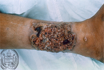
Fig.34-A1
Chromoblstomycosis
The extensive tumor-like lesions with ulcerations on the lower leg do not suggest a fungal infection.
|
|
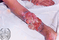
Fig.34-B1
Carcinoma
This tumor developed in a chronic ulcer of the skin. There is some similarity with the tumor shown in fig. 34-A1.
|
|
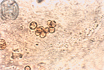
Fig.34-A2
Chromoblastomycosis
In a smear taken from the ulcer shown in fig. 34-A1 appear a cluster of fungal cells with brown capsules.
|
|
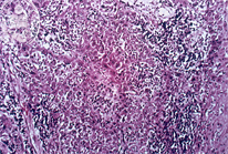
Fig.34-B2
Carcinoma
Histologically a cell-rich squamous cell carcinoma was confirmed.
|
|
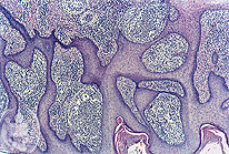
Fig.34-A3
Chromoblastomycosis
In the non ulcerated region of the skin microscopically an extensive pseudoepitheliomatous hyperplasia of the epidermis and the chronic inflammation in the dermis can be observed in this HE stain at low power. Fungus cells can not be detected in this slide.
|
|
|
|
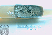
Fig.34-A4
Chromoblastomycosis
The macroscopical aspect of the fungal species of the genus Hormodendrum in a culture colony, one of the fungi which may produce this infection.
|
|
|
| español | english | deutsch |
|
|