|
XX.
Coccidioidomycosis
|
|
Case 70:
Coccidioidomycosis / Eccrine poroma
|
|
|
|
Coccidioidomycosis
|
|
Eccrine poroma
|
|
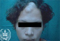
Fig.70-A1
Coccidioidomycosis
A 19-years old female patient from Barinitas in the State Barinas / Venezuela presents a hen´s egg-like skin tumor in the centre of her forehead below the hair of the head which is slightly fluctuant. The patient lost weight permanently in the last time had a heavy cough and produces abundant partly haemorrhagic mucus. In addition, a large abscess developed in the subscapular region of the back.
|
|
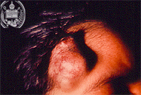
Fig.70-B1
Eccrine poroma
A 32-year old man is sent from his country doctor in Curbati to the University Hospital in Barinas. The patient has a tumor on the right side of his forehead which did grow steadily during one year, but is completely painless. Now the tumor has reached the size of a hen s egg and is located below the hairline. Superficially numerous hyperaemic vessels and small haemorrhages are seen.
|
|
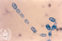
Fig.70-A2
Coccidioidomycosis
The patient is a native of the State Falcon / Venezuela. There are sandy deserts and dunes, so called "médanos", the habitat of the dimorphic fungus Coccidioides immitis. Its mycelial form grows in the sand and in the cultures at room temperatures as hyphae with arthrospores which we illustrate here. The pulmonary infection of the patient certainly took place in that region, because only in this region occur such fungal infections in our country. Now a dissemination of this fungus infection must have occurred.
|
|
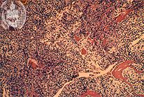
Fig.70-B2
Eccrine poroma
In the histological examination of a biopsy taken from the tumors solid parts cubic cells are found. They are basophilic and have roundish nuclei. In addition, oedema, haemorrhages and lymphatic cells are present. Several fissural cavities are lined by cubic epithelial cells and contain an acidophilic liquid. There are no signs of malignancy.
|
|
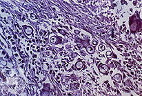
Fig.70-A3
Coccidioidomycosis
The fungus is confirmed in tissue biopsies in his parasitic form and in two types, the immature spherules, looking like yeast cells, but without buds present in this Figure. HE stain.
|
|
|
|
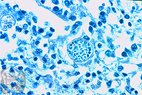
Fig.70-A4
Coccidioidomycosis
The mature types of this fungus appear in tissues as large cyst-like cells with endospores. The latter develop later after leaving the large spherules to become immature spherules.
|
|
|
|
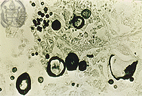
Fig.70-A5
Coccidioidomycosis
Here immature and mature parasitic cells of C.immitis are stained black with the Grocott method. The mature ones, partly destroyed, are discharging the endospores.
|
|
|
| español | english | deutsch |
|
|