|
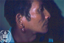
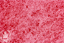
Fig.12-A1
Paracoccidioidomycosis
A 34 -year old patient from the environs of the town Barinas / Venezuela noted a subcutaneous nodule on the right jaw which had appeared several weeks ago. A biopsy was taken and the nodule was completely removed.
|
|
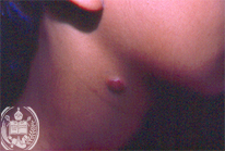
Fig.12-B1
Xanthomatous dermatofibroma
A five-year old boy from Barinas was developing a slowly growing subcutaneous tumor in the right submandibular region. It had a diameter of 1 cm and is hard and painless. The biopsy allowed the histological diagnosis.
|
|
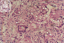
Fig.12-A2
Paracoccidioidomycosis
The resected nodule was a lymph node containing foci of destruction. In the HE stain numerous round, vacuolated yeast cells and giant cells were seen. The yeast cells were Grocott positive and showed numerous fungus cells with multiple budding, pathgnonomic for this fungal infection. In the x-rays of the thorax numerous foci were present in both lungs. The therapy with Amphotericin B was successful.
|
|
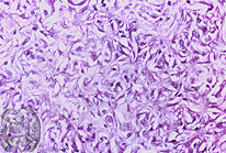
Fig.12-B2
Xanthomatous dermatofibroma
A pale colored connective tissue was found in the biopsy and some vacuoles were detected without signs of malignancy.
|