|
XI.
Chagas Disease
|
|
Case 60:
Chagas Disease / Haemangioma
|
|
|
|
Chagas Disease
|
|
Haemangioma
|
|
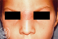
Fig.60-A1
Chagas Disease
A 9-year old boy from a village south of the town Barinas / Venezuela is suffering fever since 10 days, feels weak and has arthralgias. Above the upper parts of his nose a reddish, painful region of the skin had developed with a diameter of 2,5 cm. The fact, that the boy came from an endemic region of the Chagas disease was the reason to make a special examination of his blood which confirmed the presence of parasites of the protozoan species Trypanosoma cruzi. Therefore a so called chagoma was assumed to be present produced by the sting of a vector. Another consequence of a primary infection of this kind is the Romaña sign which we will describe in the next case.
|
|
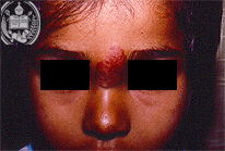
Fig.60-B1
Haemangioma
On the same place as the chagoma of fig. 60-A1 in an other patient a tumor of this kind is present.
|
|

Fig.60-A2
Chagas Disease
In the blood smears coloured by the Giemsa stain und under high power a flagellate protozoon, Trypanosoma cruzi, could be demonstrated. At his front end the nucleus and on his posterior parts an elongated cariosoma are seen. From the latter the flagellum is starting.
|
|
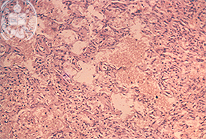
Fig.60-B2
Haemangioma
Histologically the clinical diagnosis can be confirmed. Neither inflammation, nor malignancy is found.
|
|
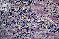
Fig.60-A3
Chagas Disease
The Chagas-infection produces frecuently a myocarditis, here also with a gigant cell. Parasites are not present in this field.
|
|
|
|
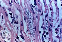
Fig.60-A4
Chagas Disease
Occasionally an elongated kinetoplasst can be recognized in an amastigote of an nest in a myocardial fiber.
|
|
|
|

Fig.60-A5
Chagas Disease
In addition to the infestation of the myocard a chagasic encephalitis is relatively frequent. Here a nest of amastigotes of Trypanosoma cruzi is demonstrated in the brain. The other organs are infested more seldom.
|
|
|
|
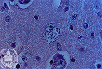
Fig.60-A6
Chagas Disease
In cases of chronic Chagas myocarditis, as in this case, giant cells can be seen, but mostly no parasites.
|
|
|
| español | english | deutsch |
|
|