|
IV.
Chromoblastomycosis
|
|
Case 30:
Chromoblastomycosis / Basalioma
|
|
|
|
Chromoblastomycosis
|
|
Basalioma
|
|
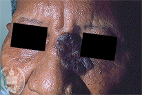
Fig.30-A1
Chromoblastomycosis
A 76-year old patient from the State Apure/ Venezuela refers that the dark tumor in his face grew slowly for several years. It was considered a birth mark, was painless and not itching.
|
|
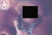
Fig.30-B1
Basalioma
In the same region as seen in fig 30-A1 a tumor is found showing ulceration and dark pigmentation.
|
|
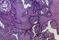
Fig.30-A2
Chromoblastomycosis
The biopsy shows a pseudoepitheliomatous hyperplasia of the epidermis and a non-specific inflammation with giant cells and microabscesses. Fungal cells are very difficult to see at this magnification and in this HE stained slide.
|
|
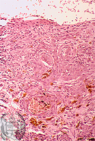
Fig.30-B2
Basalioma
The pigmentation may be detected clearly with the microscope.
|
|
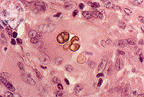
Fig.30-A3
Chromoblastomycosis
With higher power small clusters of fungus cells were observed which had a brownish capsule. The multiplication of these fungi in tissues occurs by septation and not by budding as it is seen in the yeast-like cells in other mycoses. Septa are not well seen in this.
|
|
|
| español | english | deutsch |
|
|