|
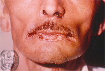
Fig.18-A1
Paracoccidioidomycosis
A 56-years old farmer from Altamira, a village in the deeper layers of the Venezuelan Andes is showing several ulcers on the lips and the left nostril, resistant to any therapy. Furthermore, the patient has ulcerations in the oral mucosa. The x-ray examination of the thorax reveals numerous focal lesions in both lungs.
|
|
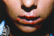
Fig.18-B1
Herpes labialis and oralis
These ulcerated, swollen and painful lips were seen in a young man who had also fever. The patient had these blisters several times and related that this affection is very painful inhibiting him to eat and drink.
|
|

Fig.18-A2
Paracoccidioidomycosis
A biopsy taken from the oral mucosa presented a non-specific granulation tissue with numerous giant cells. With lower power and the HE stain only some fungus -like suspicious elements could be seen.
|
|
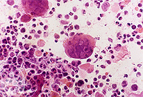
Fig.18-B2
Herpes labialis and oralis
In smears from the ground of an ulcer inflammatory cells are seen in addition to the so called "balloon cells" with inclusion bodies. Fungi or parasites were not found.
|
|
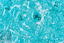
Fig.18-A3
Paracoccidioidomycosis
The yeast-like cells of the dimorphic Paracoccidioides brasiliensis multiply in the tissue by the characteristic multiple budding, which allows a reliable diagnosis. The black stained fungus cells are good to observe in the present picture, situated in a giant cell. Grocott Method.
|
|
|