|
XVII.
Phaeohyphomycosis
|
|
Case 67:
Phaeohyphomycosis / Eczema vaccinatum
|
|
|
|
Phaeohyphomycosis
|
|
Eczema vaccinatum
|
|
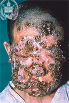
Fig.67-A1
Phaeohyphomycosis
A young man from the Venezuelan Andes is presenting some dark foci in his face and neck which formed in several months. They increased in numbers, got larger and were covered by crusts.
|
|
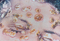
Fig.67-B1
Eczema vaccinatum
This patient presents an eczematous lesion especially on his forehead. In the periphery of the inflammatory foci a red skin is noted.
|
|
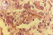
Fig.67-A2
Phaeohyphomycosis
The biopsy of one of these foci showed in the HE stain a non-specific inflammation with the presence of yellowish coloured septated hyphae.
|
|
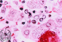
Fig.67-B2
Eczema vaccinatum
Smears of the pathologic lesions present a multinuclear epithelial giant cell and leucocytes. In several epithelial cells inclusion bodies are found surrounded by a thin halo. These are the so called Guarneri bodies.
|
|
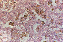
Fig.67-A3
Phaeohyphomycosis
In other fields necrotic foci are seen in the HE stain. In the field of this figure we could photograph, preferently roundish yellowish coloured elements can be seen. Apparently they are transversally cut hyphae. HE stain.
|
|
|
|
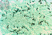
Fig.67-A4
Phaeohyphomycosis
With low power and in the Grocott stained slide numerous black coloured hyphae are present, that means fungal elements. The fungus cells with a proper colour belong to the so called group of dematiaceous fungi.
|
|
|
| español | english | deutsch |
|
|