|
XVI.
Lobomycosis
|
|
Case 66:
Lobomycosis / Lymphocytoma
|
|
|
|
Lobomycosis
|
|
Lymphocytoma
|
|
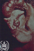
Fig.66-A1
Lobomycosis
The external ear is enlarged and thickened and has a firm consistency, this means that a typical so called keloid is present.
|
|
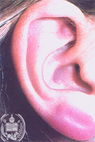
Fig.66-B1
Lymphocytoma
The external ear is thickened similar to the keloid of Fig. 66-A1. The lymphocytoma is called also "benign lymphomatosis cutis". There is a weak, often blue-red and benign tumor-like swelling already noted in childhood and young people, possibly caused by insect bites.
|
|
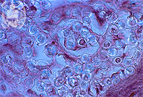
Fig.66-A2
Lobomycosis
Microscopically a dense infiltration with roundish fungus cells is seen in the dermis. These are fungus cells of the species Loboa loboi. HE stain.
|
|
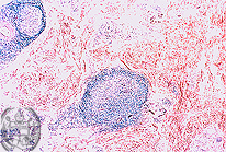
Fig.66-B2
Lymphocytoma
Histologically perivascular, round cell infiltrates are present in the deeper parts of the dermis and in the subcutaneous fat tissue, preferently consisting of lymphocytes and histiocytes. There are nodules with germinative centres. HE stain.
|
|

Fig.66-A3
Lobomycosis
The fungus cells of Loboa loboi stain black with the special staining method, i.e the GMS method.
|
|
|
|

Fig.66-A4
Lobomycosis
Some fungal cells show a birefringence when observed with the polarized light. GMS stain.
|
|
|
| español | english | deutsch |
|
|