|
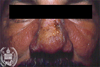
Fig.7-A1
Mucocutaneous Leishmaniasis
This 41-years old patient lives in the South of the State Bolivar, a typical tropical region. He refers that he noted a pimple on his nose some months ago which spread slowly over the back of the nose and both cheeks forming a butterfly-like figure.
|
|
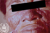
Fig.7-B1
Chronic discoid lupus erythematodes
In a 43-years old farmer wife, living in the State of Barinas existed since 6 years a blue-red, scaling discoloration of the skin, extending from the back of the nose to both cheeks.
|
|

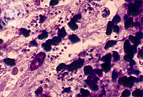
Fig.7-A2
Mucocutaneous Leishmaniasis
In the biopsy foci of histiocytes with a vacuolated cytoplasm were found containing numerous small parasites. The Grocott stain was negative.
|
|
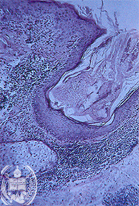
Fig.7-B2
Chronic discoid lupus erythematodes
The histological examination of the skin biopsy shows a diffuse and follicular hyperkeratosis of the epidermis. In the dermis round cell infiltrates are observed. The patient is feeling well. The treatment was successful.
|