VII. Histoplasmosis
Case 48. Histoplasmosis / Epithelioma
View Thumbnails
Fig. 48-A3
Previus
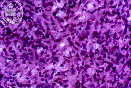
Histologically the biopsy of the ulcer on the cheek (48-A1) showed a granulation tissue with numerous histiocytes, lymphocytes and plasmacells. The few fungus cells were difficult to recognize in the HE stain and with a magnification of 400 x.
Next
| español | english | deutsch |
Facultad de Medicina Universidad de Los Andes Merida - Venezuela