|
VI.
Sporotrichosis
|
|
Case 45:
Sporotrichosis / Basalioma
|
|
|
|
Sporotrichosis
|
|
Basalioma
|
|

Fig.45-A1
Sporotrichsosis
Extensive skin ulcer of the lower leg above the angle joint of the toes with distinct tumor-like nodules in this patient.
|
|
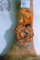
Fig.45-B1
Basalioma
On the lower leg of this patient an ulcerated tumor looking similar to a fungal infection.
|
|
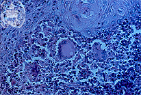
Fig.45-A2
Sporotrichosis
Typical tissue reaction in the dermis with a non-specific inflammation and several giant cells. As in this fungi mostly are not detectable, at least not in chronic cases. HE stain.
|
|
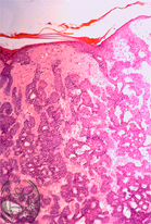
Fig.45-B2
Basalioma
Histologically an adenoid basalioma is found.
|
|
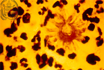
Fig.45-A3
Sporotrichosis
In an abscess of the dermis a typical asteroid body is found. These elements are occasionally present in lesions of this fungal infection. In the centre frequently a fungus cell can be recognized. In other mycoses they are not so easily found as in the sporotrichosis. HE stain; high power.
|
|
|
| español | english | deutsch |
|
|