|

Fig.38-A1
Lepromatous Lepra
A 14-years old boy presented warty and nodular skin lesions on both sides of his nose. Furthermore, an abscess in the nasal cavity and a skin thickening above the navel were noted with a diminution of the sensibility.
|
|
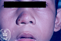
Fig.38-B1
Sebaceous adenoma
A 6-year old boy from the town of Barinas / Venezuela showed several slightly elevated spots on the skin of his face, particularly on his nose, which had developed a long time ago. They were neither painful nor itching.
|
|
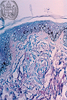
Fig.38-A2
Lepromatous Lepra
A biopsy taken from the skin lesions of the nose showed a suspicious granulation tissue. With the Ziehl-Neelsen stain some red coloured lepra bacilli were detected.
|
|
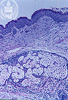
Fig.38-B2
Sebaceous adenoma
The biopsy revealed a fibrosis of the corium with the presence of partly atrophic sebaceous glands. The boy died 4 years later. The autopsy confirmed the clinical diagnosis of a tuberous brain sclerosis.
|