|
II.
Paracoccidioidomycosis
|
|
Case 15:
Paracoccidioidomycosis / Carcinoma
|
|
|
|
Paracoccidioidomycosis
|
|
Carcinoma
|
|

Fig.15-A1
Paracoccidioidomycosis
A 45-years old worker from the vicinity of the town Barinas / Venezuela complains for several months about toothache and ulceration at the right side of his tongue, which is growing continuously. Furthermore, some enlarged lymph nodes are palpable at the lower yaw border. The tongue is thickened and the ulcer is hardly seen.
|
|
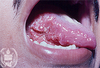
Fig.15-B1
Carcinoma
A 22 -year old man presents a painless ulcer at the right side of his tongue growing slowly. All kinds of treatment were unsuccessful.
|
|
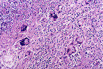
Fig.15-A2
Paracoccidioidomycosis
A biopsy taken from the border of the ulcer of the tongue shows a cell-rich granulation tissue with giant cells containing roundish, optically empty elements. HE stain, low power magnification.
|
|
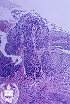
Fig.15-B2
Carcinoma
The histological examination of a biopsy from the border of the tongue ulcer revealed a non-cornified squamous cell carcinoma.
|
|
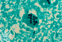
Fig.15-A3
Paracoccidioidomycosis
The round, optically empty elements turn out to be typical yeast cells of Paracoccidioides brasiliensis partly with multiple budding. GMS stain.
|
|
|
| español | english | deutsch |
|
|