|
XIV.
Amebiasis
|
|
Case 64:
Amebiasis / Carcinoma
|
|
|
|
Amebiasis
|
|
Carcinoma
|
|
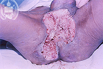
Fig.64-A1
Amebiasis
In this patient from Mérida / Venezuela developed slowly an extensive ulcerated skin lesion in the perianal region extending to the scrotum and both gluteal sides. Clinically was thought in the possibility of a malignant tumor, until a biopsy revealed the parasitic nature of an infection which could be treated successfuly with a complete recovery, showing extensive scars.
|
|
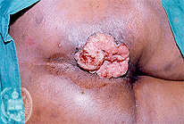
Fig.64-B1
Carcinoma
Here the tumor is shown in an early stage, but the tumor nature of this nodule exists without any doubts. The perianal region is affected as in the patient of the fig. 64-A1.
|
|
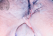
Fig.64-A2
Amebiasis
The specific treatment after the biopsy diagnosis was successful after several months. Here the extensive cicatrices are seen.
|
|
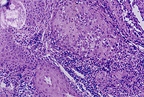
Fig.64-B2
Carcinoma
Histologically a squamous cell carcinoma was diagnosed.
|
|
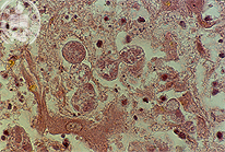
Fig.64-A3
Amebiasis
With the HE stain the relatively large intestinal amebae (Ameba histolytica) may be confused with large histiocytes.Therefore it is recommended to untrained observers to use a special staining method. Here we show the amebae in the HE stain under high power.
|
|
|
|

Fig.64-A4
Amebiasis
The Ameba histolytica, the intestinal one, stains well with the PAS method and is illustrated here in great numbers situated in a blood vessel. At the contrary, other species of amebae are PAS negative.
|
|
|
|

Fig.64-A5
Amebiasis
The application of the Grocott method is recommended especially for the examination of liver slides. This picture shows amebae at low power in the liver.
|
|
|
| español | english | deutsch |
|
|