|
XIII.
Mucormycosis
|
|
Case 63:
Mucormycosis / Basalioma
|
|
|
|
Mucormycosis
|
|
Basalioma
|
|
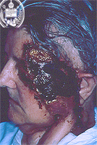
Fig.63-A1
Mucormycosis
In this patient mostly the left half of the face is occupied by a heavily pigmented inflammatory tissue.
|
|
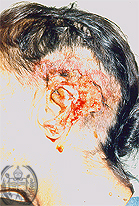
Fig.63-B1
Basalioma
As in fig. 63-A1 exists in this patient a complete destruction of an ear and the surrounding skin area by this tumor.
|
|
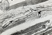
Fig.63-A2
Mucormycosis
The large branching septated hypha is good to recognize in the HE stain.
|
|
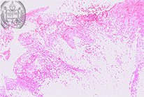
Fig.63-B2
Basalioma
The diagnosis was confirmed by histology.
|
|

Fig.63-A3
Mucormycosis
The entire lumen of this blood vessel is filled up by large hyphae stained black by the Grocott method. The invasion of blood vessels by the hyphae can be observed especially in cases of mucormycosis.
|
|
|
|
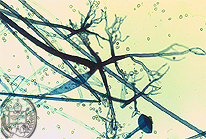
Fig.63-A4
Mucormycosis
Smear of the culture colony of Rhizopus sp. This fungus is one the many fungal species which may cause this mycosis. The rhizoid aspect of the hyphae is typical. The principal genera whose species may cause this mycosis are Rhizopus, Absidia and Mucor.
|
|
|
| español | english | deutsch |
|
|