|
X.
Rinoescleroma
|
|
Case 59:
Rinoescleroma / Carcinoma
|
|
|
|
Rinoescleroma
|
|
Carcinoma
|
|
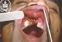
Fig.59-A1
Rhinoscleroma
In the oral cavity an ulcerative, tumorous lesion with haemorrhages in the mucosa can be reognized. Furthermore, the set of teeth is defect. On the palate a firm tumor is present. The patient declares to have noted the tumor already years ago.
|
|
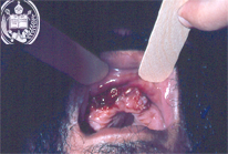
Fig.59-B1
Carcinoma
In the oral cavity of this 50-year old patient exist ulcerated papillomatous proliferations. He noted his disease 4 months ago.
|
|
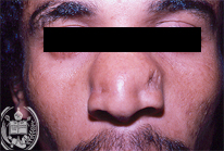
Fig.59-A2
Rhinoscleroma
In addition, the patient presents an enlargement and deformation of his nose and the aspect of a so called "tapir nose".
|
|
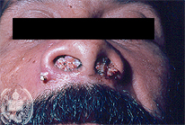
Fig.59-B2
Carcinoma
Also in the nasal cavities and outside the nose tumorous tissues and nodules are present.
|
|
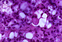
Fig.59-A3
Rhinoscleroma
In the inflammatory tissue of the oral cavity typical histiocytes are present, called Mikulicz cells and a so called Russel body. HE and high power.
|
|
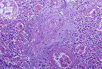
Fig.59-B3
Carcinoma
Histologically slightly cornified squamous cell carcinoma was diagnosed.
|
| español | english | deutsch |
|
|