|
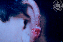
Fig.1-A1
Mucocutaneous Leishmaniasis
A 27 years-old vegetable seller from Barinas, a town at the foot of the Venezuelan Andes, develops within several weeks a very itching, papulo-erythematous, ulcerated tumor on the helix of the left ear. The borders of the tumor are lightly elevated and are bleeding easily when touched. In its surrounding the skin is covered by a brownish crust.
|
|
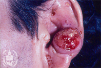
Fig.1-B1
Epithelioma
A 41-year old man from Barinas comes to a dermatologist, presenting a tumor on his left ear, which grew steadily during the past 5 months. The tumor has a diameter of 20 mm, is round and elevated, very painful and secrets a serous -hemorrhagic fluid. In the cervical and sub maxillary region some painless enlarged lymphnodes are felt. A biopsy is taken from the border of the tumor.
|
|
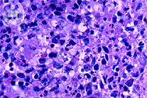
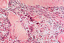
Fig.1-A2
Mucocutaneous Leishmaniasis
The histology of the specimen taken from the border of the ulcer shows a certain amount of histiocytes with foamy cytoplasm, accompanied by plasma- and lymphocytes. Within the cytoplasm of the histiocytes parasites from the type Leishmania brasiliensis are seen. Several histiocytes show an optically empty centre, whereas the parasites lay in the periphery. This effect occurs during the mechanical preparation and coloration of the slides.
|
|
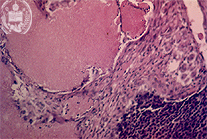
Fig.1-B2
Epithelioma
The histological examination of the specimen shows a benign calcifying epithelioma (Malherbe). It consists of typical basophilic matrix cells surrounded by connective tissue and acidophilic "shadow cells".
|